Bitewing X-Ray: Definition, Uses & Key Differences
Bitewing x-ray is a common dental imaging technique used to detect tooth decay, bone loss, and other potential issues between teeth. In this technique, the patient gently bites down on a special tab that holds the x-ray sensor or film in place. This allows the upper and lower teeth to be captured in a single image.
Dentists use a bitewing x ray machine to examine molars and premolars closely. Compared to traditional x-rays, digital bitewing x ray provides faster imaging with reduced radiation exposure, making it a safer and more efficient option for both patients and dental professionals.
The dental x ray bitewing method is particularly effective for identifying cavities, bone loss, and issues with dental restorations. With its ability to detect problems in early stages, bitewing x ray dental remains an essential tool in modern dentistry.
What Is a Bitewing X-Ray?
The bitewing x-ray definition refers to a type of dental radiograph that captures both the upper and lower teeth in one image to detect cavities, bone loss, and other dental concerns. This technique is commonly used in routine dental exams.
Using a bitewing x ray machine, dentists can closely examine the oral structures for signs of decay or gum disease. The procedure involves the patient biting down on a film or digital sensor, which is positioned to provide a clear view of the teeth's contact points.
Modern practices often prefer digital bitewing x ray systems for their improved accuracy, faster results, and lower radiation exposure. As a result, bitewing x ray dental is considered a vital part of preventive dental care.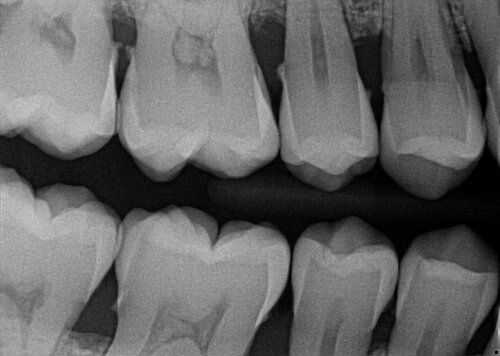
Why Are Bitewing X-Rays Used in Dentistry?
Bitewing x-ray imaging plays a crucial role in modern dentistry by enabling early diagnosis of dental issues. Key benefits of this technique include:
- Early Cavity Detection: Bitewing dental x ray is highly sensitive in detecting cavities between teeth, even in areas that are difficult to spot during a visual examination.
- Gum Disease Diagnosis: Bitewing x-ray dental helps identify bone loss and signs of periodontal disease below the gum line.
- Assessment of Fillings and Restorations: Dentists use bitewing x ray dental images to ensure dental fillings, crowns, and other restorations are properly fitted and intact.
- Monitoring Oral Health Changes: By comparing past and present x ray bitewing images, dentists can track changes in oral health over time.
Do Dentists Still Use Bitewing X-Rays?
Yes, dentists continue to use bitewing x-ray imaging as a reliable diagnostic tool. Despite technological advancements, this method remains essential for identifying dental issues that may go unnoticed during visual examinations.
Key reasons for its continued use include:
- High Accuracy and Detail: X ray bitewing captures fine details between teeth, providing clear images for cavity detection.
- Low Radiation Exposure: Digital bitewing x ray systems emit minimal radiation, enhancing patient safety.
- Quick and Efficient Results: Digital systems deliver instant images, enabling faster diagnosis and improved patient care.
While some cases may require a broader view through a panoramic x ray vs bitewing comparison, bitewing x ray dental remains the gold standard for targeted cavity detection.
How to Take a Bitewing X-Ray: Step-by-Step Guide
Performing a bitewing x ray requires precision and proper technique. Here’s a step-by-step guide for optimal results:
- Prepare the Necessary Equipment
- Bitewing x ray machine (digital or film-based)
- Protective lead apron and thyroid shield
- Disposable sensor covers and gloves
- Position the Patient Correctly
- Ensure the patient is seated upright with their head stabilized.
- Place the lead apron on the patient for radiation protection.
- Align the patient’s head parallel to the x-ray device.
- Insert the Sensor and Bite Tab
- Position the sensor to capture both upper and lower teeth.
- Instruct the patient to bite down gently on the special bite tab.
- Position the X-Ray Machine
- Align the x-ray beam at the correct angle to capture the desired area.
- Ensure the device is positioned to clearly visualize the contact points between teeth.
- Capture the X-Ray Image
- Activate the x-ray beam briefly while the operator stands behind a protective barrier.
- For digital bitewing x ray, the image appears instantly; for film-based x-rays, chemical processing may be required.
- Evaluate the Image
- Assess the image for clarity, ensuring cavities, bone loss, and other concerns are visible.
- If the image is unclear, adjustments may be necessary.
- Communicate Findings to the Patient
- Explain the results and discuss possible treatments.
- Schedule follow-up appointments if needed.
Bitewing X-Ray vs. Panoramic X-Ray: Key Differences
|
Feature |
Bitewing X-Ray |
Panoramic X-Ray |
|
Detailed Cavity Detection |
✔️ Yes |
❌ Limited |
|
Gum Disease and Bone Loss |
✔️ Yes |
✔️ Yes |
|
Orthodontic Planning |
❌ No |
✔️ Yes |
|
Impacted Tooth Detection |
❌ No |
✔️ Yes |
While bitewing x ray dental is ideal for detailed cavity detection, panoramic x ray provides a broader view for orthodontic planning and impacted teeth detection.
Best AI Solutions for Bitewing X-Ray Interpretation
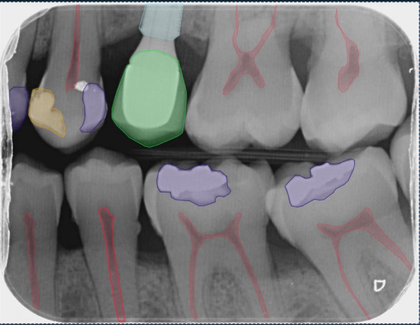
AI technology has revolutionized dental diagnostics, offering faster and more accurate bitewing x-ray interpretation. Advanced AI for dental systems provide several key benefits:
✅ Fast and Accurate Diagnoses: AI identifies cavities, fractures, and bone loss with exceptional precision.
✅ Early Detection of Dental Issues: AI can spot small issues that may be missed during visual examination.
✅ Minimized Diagnostic Errors: Automated analysis reduces the risk of human error.
✅ Enhanced Patient Communication: Visual AI reports help patients understand their dental conditions better.
CranioCatch offers an innovative AI-powered solution that enhances bitewing x-ray analysis. With its advanced algorithms, CranioCatch efficiently identifies dental problems such as cavities, periodontal issues, and restoration defects.


 Contact Us

