Cone Beam CT Scan: Everything You Need to Know

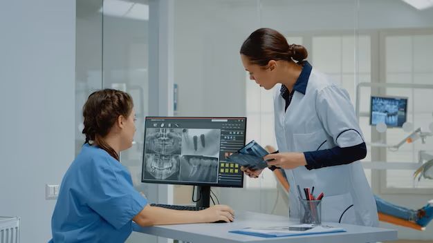
Cone beam CT scan is a specialized type of x-ray technology used by dental professionals to generate 3D images of the teeth, jaw, and surrounding structures. This cutting-edge imaging method allows for more precise diagnosis and treatment planning, particularly in complex dental procedures. Cone beam CT scan dental imaging has transformed dental care by providing detailed anatomical views that were previously unavailable. Whether it’s for implant placement, root canal treatments, or orthodontics, CT cone beam scan technology offers a comprehensive perspective of oral structures. The ability to capture high-quality images with minimal radiation exposure makes it a preferred choice for dentists today.
What is Cone Beam CT?
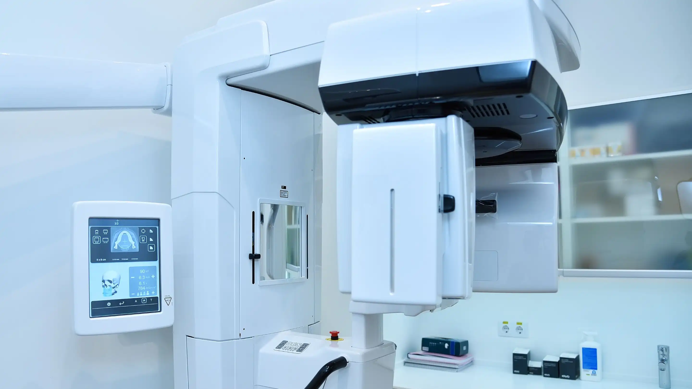
The cone beam CT is an advanced imaging technique used to generate 3D images of the teeth, jaw, and surrounding structures. This imaging method employs a cone-shaped X-ray beam that rotates around the patient’s head, capturing multiple angles in a single scan. Compared to traditional X-rays, which only produce 2D images, the CT cone beam provides a comprehensive view of the entire oral and maxillofacial region.
One of the significant applications of cone beam CT dental imaging is in diagnosing complex dental conditions. For example, dentists rely on dental cone beam CT scans to evaluate bone density, nerve positioning, and the precise structure of the jaw, which are critical factors in dental implant planning and other surgical procedures. The versatility of CT cone beam dental technology makes it an indispensable tool for modern dental practices.
In addition to its diagnostic capabilities, the cone beam CT scan dental imaging system significantly reduces radiation exposure compared to traditional CT scans. This combination of safety and precision explains why the technology is increasingly popular in dentistry.
How Does Cone Beam CT Work?
The cone beam CT scan process is both efficient and highly effective. Unlike traditional imaging techniques, this advanced system captures detailed 3D images in a single rotation around the patient’s head, ensuring high-quality visuals with minimal discomfort. Below is a step-by-step breakdown of how the CT cone beam scan operates:
• Rotation Around the Patient: The scanner rotates 360 degrees, capturing multiple images from different angles.
• Cone-Shaped X-Ray Beam: The unique cone-shaped beam covers a wider area, allowing for a comprehensive scan in one rotation.
• Image Reconstruction: Advanced software processes the captured images to reconstruct a detailed 3D model of the teeth, jaw, and surrounding structures.
• Reduced Radiation Exposure: Compared to traditional CT scans, the CT cone beam dental system minimizes radiation while maintaining image clarity.
This efficient process ensures that patients receive accurate diagnoses and customized treatment plans. Whether for implants, orthodontics, or complex surgeries, the cone beam CT scan dental system is a game-changer for dental professionals.
Why is Cone Beam CT Used in Dentistry?
The cone beam CT scan is a transformative tool in dentistry due to its ability to provide highly detailed 3D images of oral and maxillofacial structures. This advanced imaging technology is primarily used for precise diagnosis and treatment planning in complex cases, such as dental implants, orthodontics, and root canal therapies. Unlike traditional X-rays, the CT cone beam dental system captures both hard and soft tissues, offering a comprehensive view of the patient’s anatomy.
Dentists rely on cone beam CT dental imaging to assess bone density, nerve pathways, and the alignment of teeth and jaws. For example, when planning dental implant placement, the dental cone beam CT scan ensures accurate positioning by mapping the jaw’s intricate structure. Similarly, it aids in diagnosing TMJ disorders and impacted teeth, reducing the likelihood of complications during procedures.
With its reduced radiation exposure compared to conventional CT scans, the CT cone beam is a safer option for patients who require multiple scans. Its unparalleled accuracy and safety make the cone beam CT scan dental technology an essential component of modern dental care.
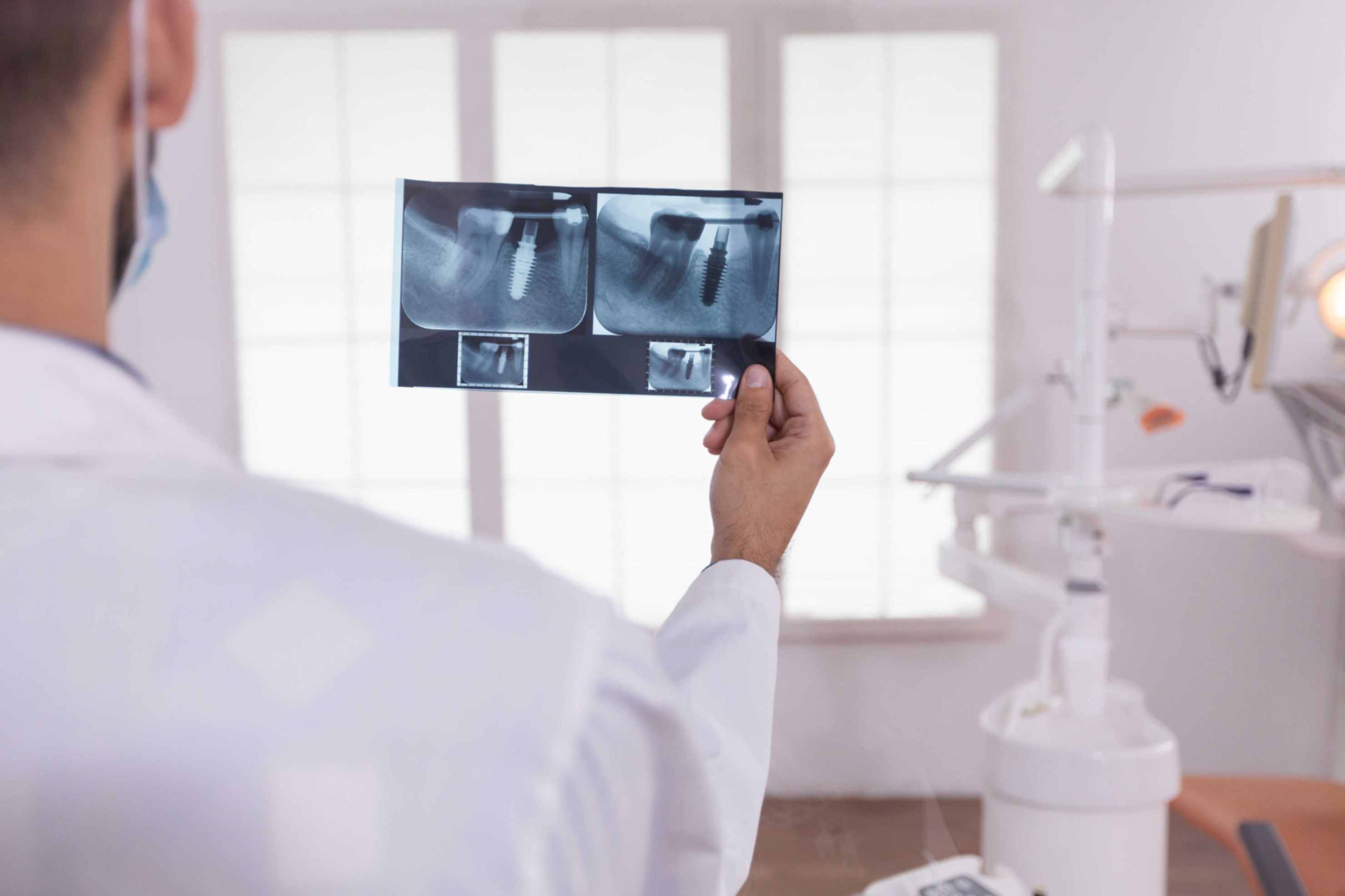
Dental Cone Beam CT for Implants and Surgeries
The cone beam CT scan is indispensable in dental implantology and surgical planning. By providing detailed images of bone quality and nerve positioning, the dental cone beam CT scan ensures precise implant placement, minimizing risks and enhancing treatment outcomes. Its ability to map the intricate anatomy of the jaw makes it a preferred tool for planning complex oral surgeries.
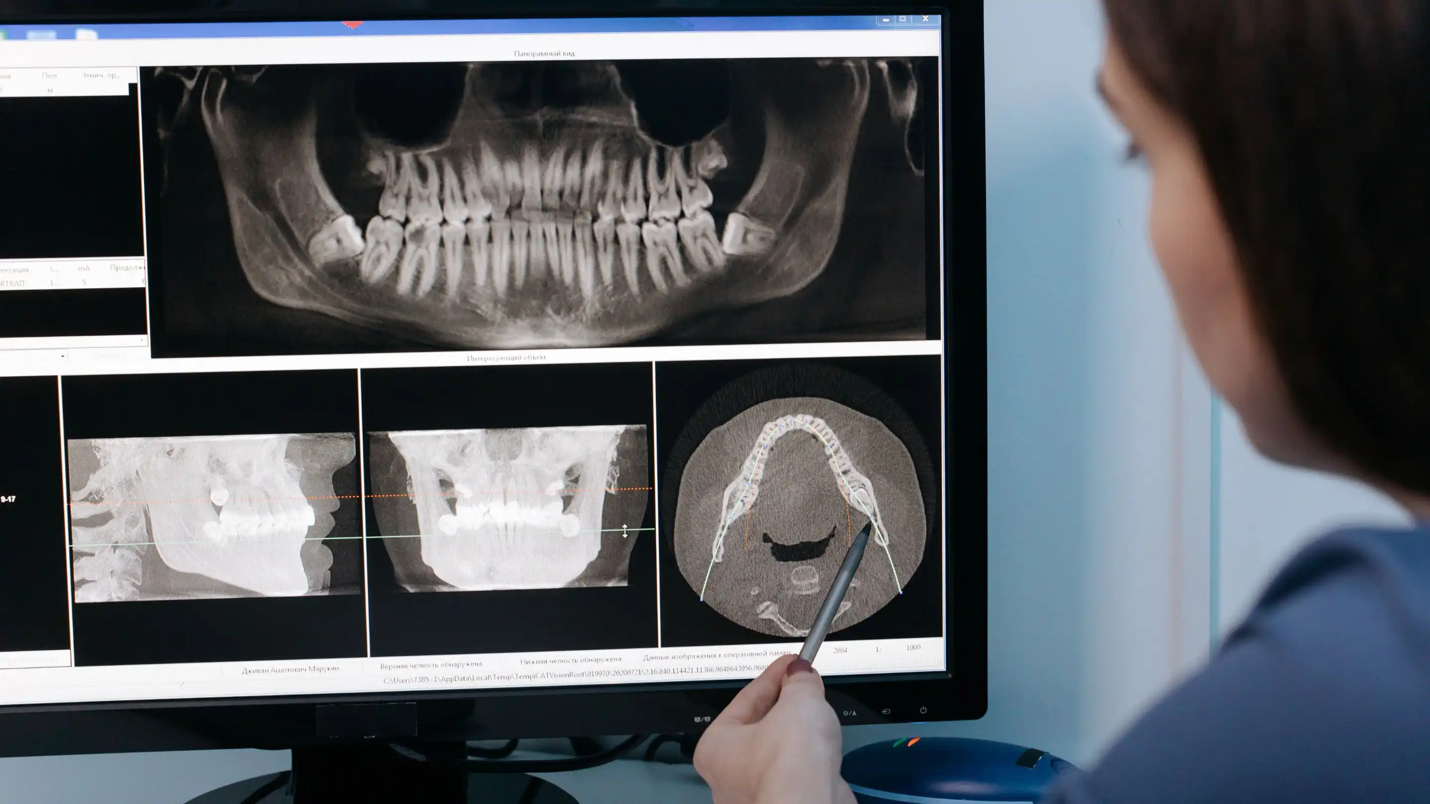
Diagnosing Dental Problems with CBCT
For diagnosing hidden dental issues, the CT cone beam dental system is unparalleled. It identifies conditions such as impacted teeth, cysts, and TMJ disorders with remarkable clarity. The cone beam CT dental technology allows dentists to detect and address problems early, improving patient outcomes.
How CranioCatch Enhances CBCT Analysis with AI
Innovative solutions like CranioCatch leverage artificial intelligence to further enhance the diagnostic potential of cone beam CT scans. By integrating AI-driven tools, CranioCatch provides dentists with precise and automated analyses of CT cone beam images. This streamlines treatment planning and improves accuracy. To learn more about AI’s role in dental imaging, visit AI in Dentistry.
How is Cone Beam CT Different from Other Imaging Techniques?
The cone beam CT scan differs from other imaging methods in both functionality and applications. Traditional dental X-rays produce 2D images, which are often limited in scope and clarity. In contrast, the CT cone beam dental system provides detailed 3D imaging, allowing dentists to examine the complete anatomy of the oral and maxillofacial region.
One of the main distinctions is the shape of the X-ray beam. The cone beam CT uses a cone-shaped beam that captures a wider area in a single scan, while traditional X-rays utilize a narrow, linear beam. This broader coverage reduces scan time and radiation exposure, making the CT cone beam scan more efficient and patient-friendly.
Additionally, the dental cone beam CT scan offers higher resolution, enabling accurate visualization of bones, nerves, and soft tissues. These capabilities make it ideal for procedures like implants and orthodontic planning, where precision is crucial. The lower radiation dose and superior diagnostic quality of the cone beam CT scan dental technology highlight its superiority over conventional imaging techniques in dentistry.
CBCT vs. Standard Dental X-Rays
While traditional X-rays are useful for detecting basic dental issues, they fall short in providing the detailed views necessary for complex treatments. The CT cone beam x-ray produces high-resolution 3D images, capturing the full scope of oral structures. This capability is particularly crucial for dental implants and orthodontics, where precise imaging is essential.
What is the Difference Between CT and Cone Beam CT?
The primary difference lies in the beam shape and application. Traditional CT scans use fan-shaped beams, while the cone beam CT employs cone-shaped beams for a broader field of view. Additionally, the cone beam CT scan dental system emits significantly lower radiation, making it safer for repeated use.
Is Cone Beam and CT Scan the Same Thing?
A cone beam CT scan and a traditional CT scan are not the same, though they share certain similarities. Both are advanced imaging technologies that provide detailed, 3D visualizations, but their applications and technical specifications differ significantly.
A traditional CT (computed tomography) scan uses a fan-shaped X-ray beam to capture multiple cross-sectional images of the body. These slices are compiled into a complete 3D view, making CT scans ideal for diagnosing a wide range of medical conditions. On the other hand, a dental cone beam CT scan uses a cone-shaped X-ray beam, which captures a focused, smaller area in greater detail. This makes it especially useful for dental and maxillofacial imaging.
One major distinction is the radiation dose. While both use X-rays, cone beam CT scans generally expose patients to lower levels of radiation compared to medical CT scans, emphasizing their safety for dental procedures. The compact size of the cone beam CT scan machine also allows for quick and convenient imaging in a clinical setting.
In summary, cone beam CT is optimized for dental and facial imaging, while traditional CT scans are designed for broader medical purposes, showcasing their unique roles in healthcare.
How Much Radiation in a Dental Cone Beam CT Scan?
The radiation exposure from a dental cone beam CT scan is relatively low compared to medical CT scans, yet higher than standard dental X-rays. Typically, a cone beam CT scan involves a dose of 29 to 477 microsieverts (µSv), depending on the machine and the area scanned. For context, this is equivalent to a few days to weeks of natural background radiation exposure.
Modern cone beam CT scan machines are designed to minimize radiation by focusing the X-ray beam only on the area of interest. They also offer adjustable settings, allowing dentists to optimize the scan for each patient’s specific needs. This ensures that the imaging process is not only precise but also safe.
It is important to note that the benefits of cone beam CT scan radiation often outweigh the risks. By providing detailed, accurate images, the technology reduces the need for repeat scans, lowering cumulative radiation exposure over time. Dental professionals evaluate the necessity of the scan to ensure patient safety, particularly for vulnerable groups like children and pregnant women
Benefits of Dental Cone Beam CT Scans
The dental cone beam CT scan offers numerous advantages for both dentists and patients:
1. Enhanced Image Quality: High-resolution 3D imaging for detailed diagnosis.
2. Accurate Treatment Planning: Ensures precise implant placement and surgical outcomes.
3. Lower Radiation Exposure: Safer for patients requiring multiple scans.
4. Comprehensive Diagnostic Tool: Visualizes both hard and soft tissues.
5. Faster Imaging Process: Reduces patient time in the chair.
These benefits underscore why the CT cone beam dental system is a cornerstone of modern dentistry.
How to Prepare for a CT Cone Beam Dental Scan
Preparing for a CT cone beam dental scan is simple and requires minimal effort:
• Remove all metallic objects, including jewelry and dental appliances, to avoid image interference.
• Inform your dentist of any medical conditions or concerns about radiation exposure.
• Stay still during the scan to ensure image clarity.
By following these steps, patients can ensure accurate and efficient imaging, contributing to better diagnostic outcomes. For clinics leveraging AI-driven analysis, check out how CranioCatch empowers dental practices:AI Dental Clinic.
How Long Does a Cone Beam CT Scan Take?
A cone beam CT scan is a quick and efficient imaging process, typically taking 30 seconds to 1 minute to complete. The process begins with the patient seated or standing, depending on the design of the cone beam CT scan machine. As the machine rotates around the patient’s head, it captures hundreds of images from various angles.
The actual scanning time is usually under a minute, but the preparation phase—such as positioning the patient and ensuring proper alignment—may add a few extra minutes. Once the scan is complete, the images are processed by specialized software to create detailed 3D visualizations of the oral and facial structures.
This efficiency makes dental cone beam CT scans ideal for busy dental practices, enabling professionals to obtain high-quality images in a matter of moments. Whether used for implant planning, orthodontics, or diagnosing complex dental issues, the speed of the scan ensures minimal disruption for patients while delivering maximum diagnostic value.

 Contact Us