Blogs

You can find current information about Craniocatch here!

Dental Digital Radiography

Dental Digital Radiography

Dental digital radiography has revolutionized the field of dentistry by offering advanced imaging techniques that provide detailed insights into oral health. One of the key components of digital dentistry is the utilization of digital x-ray machines, enabling dental professionals to capture high-quality images for diagnostic purposes.
 

Dental X-Ray Machines

When it comes to digital x-ray machines, there are various types available in the market. These machines utilize advanced technology to produce detailed images of the teeth and surrounding structures. The benefits of digital dental radiography are vast, ranging from improved image quality to reduced radiation exposure for patients.


How Does It Work Dental Radiography ?

Dental radiography is a crucial component of modern dental health care. Digital dental x-ray technology, also known as dental digital radiology, offers numerous advantages over traditional dental films. Digital x-ray for teeth uses x-ray sensors instead of dental films, resulting in less radiation exposure for patients. The x-ray image captured by the digital system can be viewed instantly on a computer screen, allowing for quick and accurate diagnosis. This digital radiography system is also beneficial for dental assistants and insurance companies, as it decreases the need for storing and managing physical dental films.

There are different types of digital dental and intraoral x-rays that can be taken using digital imaging software. The digital radiography systems are widely used in many dental practices due to their convenience and radiation safety. While traditional dental equipment used dental films that were bulkier and required chemical processing, digital dental xray technology eliminates these drawbacks, providing a clearer and more efficient x-ray image. In addition, the digital x-ray sensors used in digital imaging systems offer less radiation than film, making them a preferred type of x-ray for imaging the upper and lower teeth.
 

Uses-Why Would I Need Digital X-rays?

Using digital dental x-rays as digital images is essential in modern dentistry. Digital imaging technologies make your existing imaging tasks more efficient. CBCT imaging can be used for dental implant placement, while digital x-ray sensors are used by dental professionals to convert dental x-rays.

Digital dental x-ray for teeth is a type of direct digital dental imaging systems that utilizes phosphor plates or digital imaging and communications in medicine (DICOM) images. Film and digital imaging technologies are both used in the dental field, with digital imaging requiring less space and being less bulkier than dental films.
 

How Often Do You Need to Conduct Digital Dental Imaging?

When it comes to digital dental x rays, it is recommended to have them done regularly as part of your dental check-up routine. With the advancement of technology in dentistry, more dentists are opting for digital x ray for teeth as it provides clearer images and reduces radiation exposure. Dental imaging is crucial in detecting any oral health issues early on and determining the best course of treatment.

The types of intraoral x-rays used in dental imaging include bitewing, periapical, and occlusal x-rays, each serving a different purpose in assessing oral health. Dental practices that go digital use digital x-ray sensors to capture images, which are then stored digitally for easy access and comparison over time.

With the advancements in digital storage and technology dental, there are now more digital options available for imaging needs. This includes panoramic imaging and other forms of radiographic imaging that provide a comprehensive view of the teeth and surrounding structures. Dental hygienists also play a crucial role in using new digital imaging technology to assist dentists in diagnosing and treating oral health issues.

Types of Digital X-Ray Machines

There are different types of digital x-ray machines used in dentistry, including intraoral and extraoral systems. Intraoral x-ray machines are designed to capture detailed images of individual teeth, while extraoral machines are used for capturing broader images of the entire oral cavity.

Benefits of Digital Dental Radiography

The advantages of digital dental radiography are significant. These include faster image processing, lower radiation exposure, and enhanced image quality, allowing dentists to make more accurate diagnoses and treatment plans.

Utilizing Digital Radiography in Dentistry

Dentists across the globe are incorporating digital radiography into their practices due to its numerous benefits. Digital imaging systems provide a more efficient workflow, allowing for quick image acquisition and enhanced patient communication.
 

Digital Imaging in Dentistry

Digital imaging plays a crucial role in modern dentistry, offering dentists a clearer view of oral structures and conditions. Digital image sensors are used in dental practice to capture detailed images that aid in diagnosing dental issues and planning treatments effectively.

Advantages of Intraoral Digital Radiography

Utilizing intraoral digital radiography provides dentists with detailed images of individual teeth and specific areas of concern. These images help in detecting dental caries, periodontal issues, and other oral health problems at an early stage.

Computed Tomography Applications in Dentistry

Computed tomography (CT) has revolutionized the way dentists diagnose and treat complex dental conditions. By utilizing CT scans, dentists can obtain detailed 3D images of the teeth, jawbone, and surrounding structures, aiding in oral surgeries and implant placements.
 

Dental Radiography Benefits

One of the significant advantages of digital dental radiography is the reduced radiation exposure compared to traditional x-ray methods. This technology enhances dental implant procedures by providing detailed images for precise implant placement and integration.

Improving Diagnoses with High-Quality Digital Images

The high-quality digital images obtained through dental radiography assist dentists in making accurate diagnoses. From detecting cavities to assessing bone health, digital radiographs play a crucial role in modern dentistry.

Enhancing Dental Implant Procedures with Digital Radiography

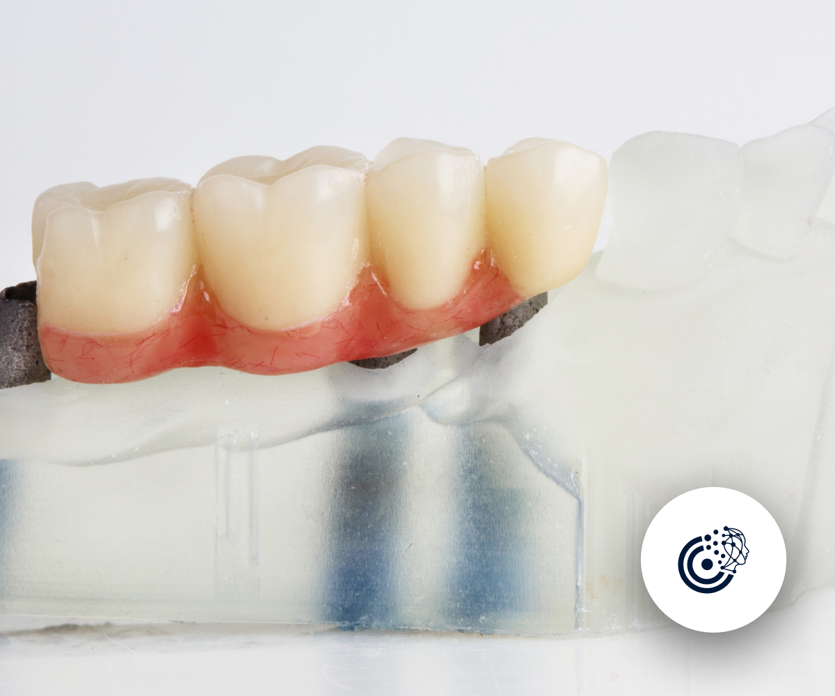
Digital radiography has significantly improved the success rate of dental implant procedures by enabling precise planning and placement. Dentists can evaluate bone density, assess the surrounding structures, and create custom treatment plans using digital images.

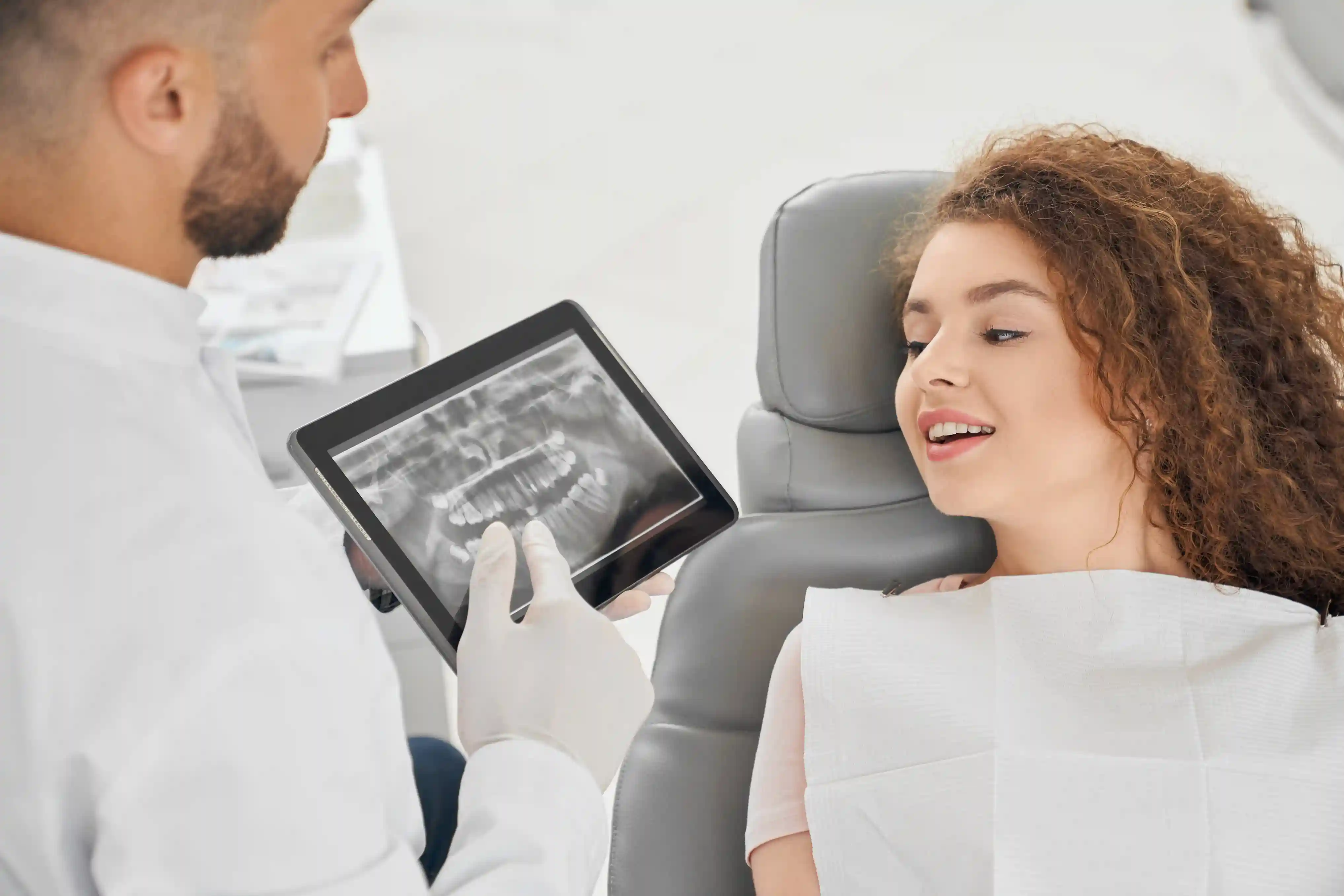
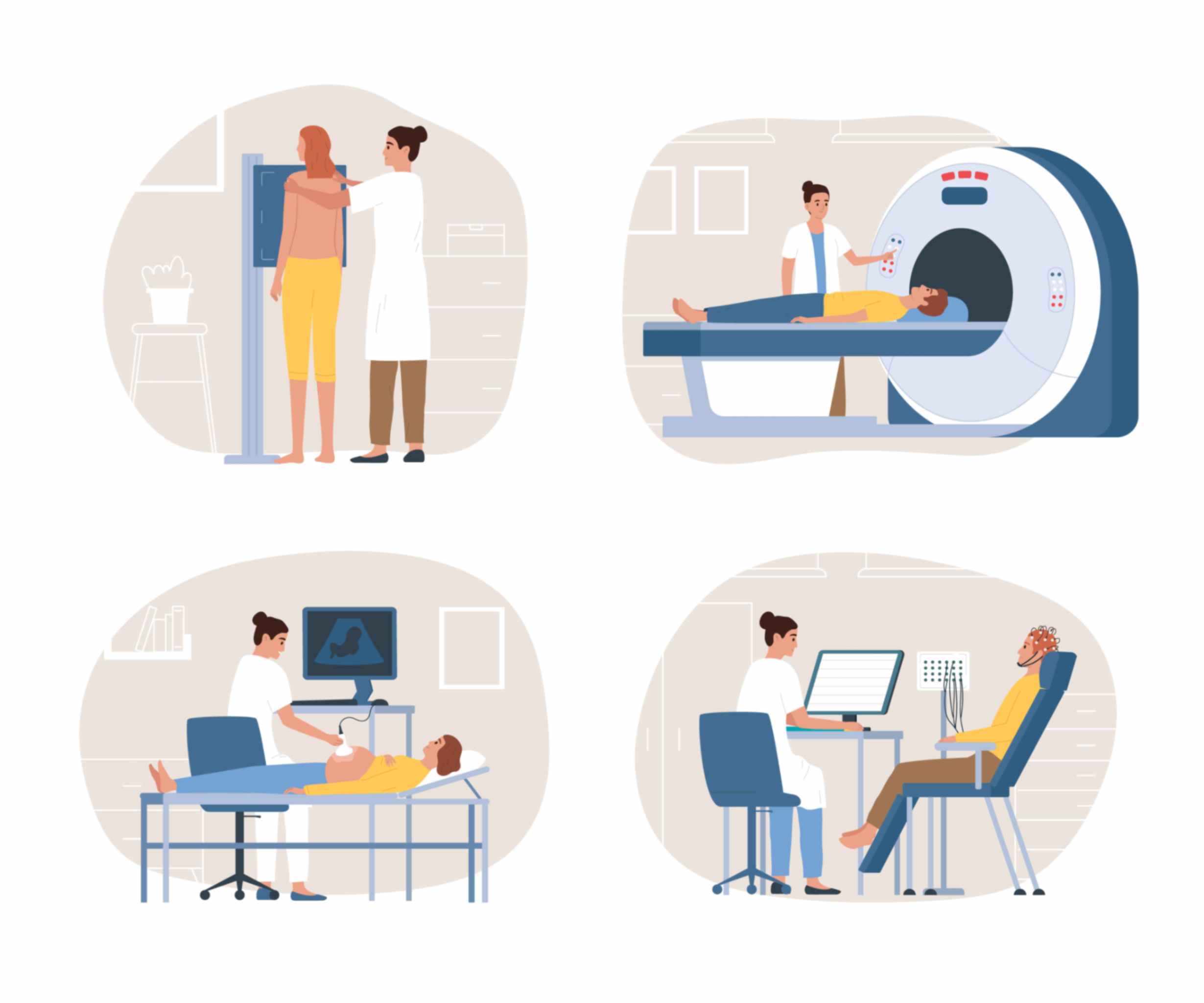
A physician examines a patient based on dental imaging results.

Evolution of Digital Dental Radiographs

The transition from traditional to digital radiography has transformed how dental imaging is performed. Incorporating digital panoramic x-rays in dental practice allows dentists to capture comprehensive images of the entire oral cavity with minimal radiation exposure.

The Role of Digital Technology in Dental Cone Beam Computed Tomography

Dental cone beam computed tomography (CBCT) has emerged as a valuable tool in modern dentistry, offering detailed 3D images for precise treatment planning. By using digital technology, dentists can visualize complex dental structures and plan surgeries with accuracy.


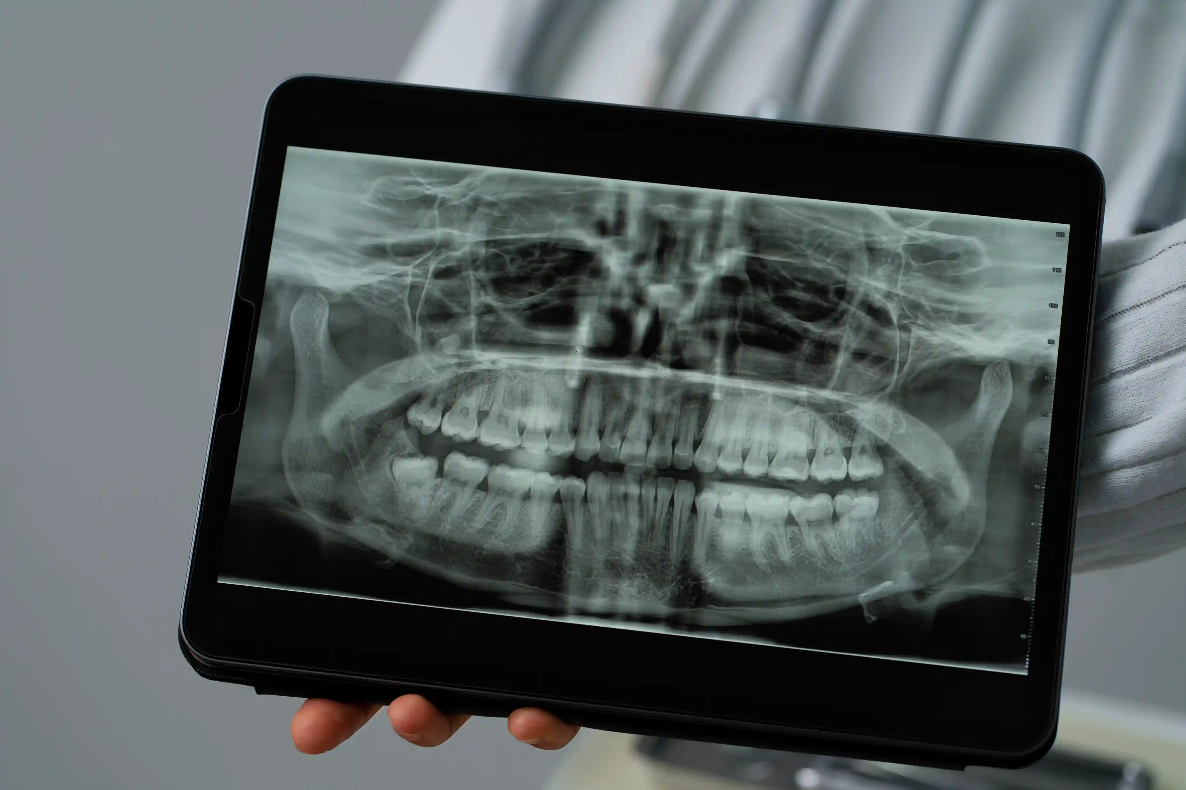
Panoramic X-rays

Incorporating Digital Panoramic X-Rays in Dental Practice

Digital panoramic x-rays provide a broad view of the oral and maxillofacial region, aiding dentists in diagnosing impacted teeth, TMJ disorders, and other oral conditions. The digital format allows for easy storage and sharing of images for consultations and referrals.

Digital Radiography Implementation

Integrating digital x-ray systems in dental clinics enhances workflow efficiency and improves patient care. Understanding the working principle of digital sensors is crucial for obtaining high-quality images and accurate diagnoses.

Maintaining Image Quality in Digital Radiographs

Ensuring consistent image quality in digital radiographs requires proper maintenance of equipment and regular calibration of imaging systems. Dentists must follow best practices to optimize image resolution and ensure accurate interpretations.

Integrating Digital X-Ray Systems in Dental Clinics

The American Dental Association recommends the use of digital x-ray systems due to their numerous benefits over traditional film-based methods. Dental clinics that implement digital radiography can enhance patient care, reduce waiting times, and streamline administrative tasks.


Cone Beam Computed Tomography with Oral Pathology

Cone Beam Computed Tomography (CBCT) is a type of dental imaging that is increasingly being used in oral pathology to provide detailed 3D images of the oral and maxillofacial regions. This technology allows for more precise diagnosis and treatment planning for conditions such as tumors, cysts, and impacted teeth.

CBCT is just one of the many types of digital imaging that is revolutionizing the field of oral pathology. By providing high-quality images with minimal radiation exposure, dental professionals are able to detect and monitor oral diseases more effectively, leading to improved patient outcomes.
 

Cone Beam Computed Tomography in Orthodontics

Cone Beam Computed Tomography is a valuable tool in orthodontics as it provides detailed 3D images of the teeth, jaw, and surrounding structures. This technology allows orthodontists to accurately assess the position of teeth and plan treatment more effectively. Dental imaging is essential in identifying any underlying issues and determining the best course of action for each patient.
 

Cone Beam Computed Tomography in Oral Surgery

Cone Beam Computed Tomography technology is widely used in oral surgery for precise imaging of the oral and maxillofacial regions. This imaging technique provides detailed 3D images of the teeth, bones, nerves, and tissues, allowing dentists to accurately plan and perform surgeries. The use of dental imaging ensures better diagnostics, treatment planning, and outcomes in oral surgery.
 

FAQ

What is digital radiography in simple terms?

Digital radiography  is a modern technique used in dentistry to capture dental images using electronic sensors instead of traditional x-ray film. This results in faster image production and clearer visuals for the dentist to analyze. Digital radiography also reduces radiation exposure for patients compared to traditional methods.
 

What are the applications of digital radiographs in dentistry ?

Dental imaging plays a crucial role in the field of dentistry. It is used to diagnose oral health issues such as cavities, gum disease, and infections. Digital radiographs provide high-quality images quickly, allowing dentists to detect problems early and create treatment plans effectively. Additionally, they reduce radiation exposure for patients.


What is the difference between direct radiography and digital radiography?

Direct radiography involves the use of a special sensor that captures x-ray images directly onto a computer screen, eliminating the need for traditional x-ray film. On the other hand, digital radiography utilizes standard x-ray machines combined with digital sensors to capture images that can be viewed and manipulated digitally.

The main difference between the two is in how the images are captured and processed. Direct radiography provides instant images, while digital radiography requires some processing before the images can be viewed. Both methods are commonly used in dental imaging to detect issues such as cavities, fractures, and abnormalities in the teeth and jaws.
 

What are the three types of digital radiography?

Dental imaging is essential in the field of dentistry for diagnosing and treating various oral conditions. There are three types of digital radiography commonly used in dental practices: intraoral, extraoral, and panoramic. Intraoral radiography involves capturing images of individual teeth and surrounding structures. Extraoral radiography captures images of larger areas like the whole mouth or jaw. Panoramic radiography provides a complete view of the entire dental structure.

Authors

CranioCatch Named Finalist at Health Tech Demo Day
May 25, 2022, 3:44 p.m.
CranioCatch at the final of "Health Technologies Demo Day"...
Inspect
Itu Core Pre-Incubation
May 25, 2022, 3:42 p.m.
CranioCatch's venture to ITU Çekirdek Kuluçka...
Inspect
AI-Powered CranioCatch Impresses at BEBKA SeedUP Demo Day!
May 25, 2022, 3:43 p.m.
CranioCatch SeedUP demo day...
Inspect
Telefon - Phone Contact Us
Beyaz Gazete icon
Milliyet icon
Trt Haber icon
Habertürk Logo
Sondakika Image