What Is a Cephalometric X-Ray? Cephalometric X-Ray vs. Standard X-Ray
Cephalometric imaging is a specialized type of x-ray technology widely used in orthodontics to evaluate and analyze the alignment of the jaw, teeth, and surrounding craniofacial structures. Unlike standard x-rays, which focus on a localized area such as a tooth or bone, cephalometric x-rays capture a broader, lateral view of the head, providing critical insights into facial symmetry, jaw relationships, and airway structure. This form of imaging plays a pivotal role in orthodontic planning and treatment, ensuring accurate diagnosis and improved patient outcomes. With advancements in digital cephalometric imaging, practitioners can now achieve greater precision and efficiency during the diagnostic process.
What Is a Cephalometric X-Ray?
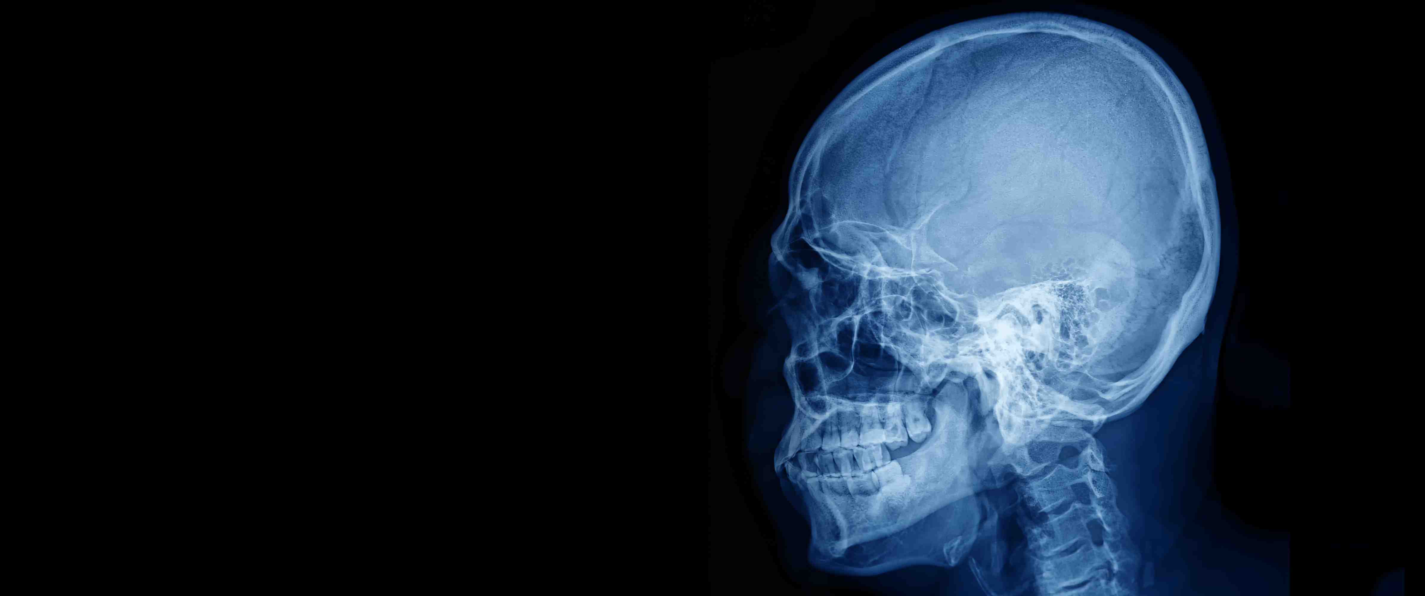
A cephalometric x-ray is a diagnostic tool primarily used in dentistry and orthodontics to create a lateral view of the patient’s head. This imaging method provides a detailed picture of the skull, jaw, and teeth, allowing practitioners to evaluate their alignment and interaction.
Unlike standard dental x-rays that focus on specific teeth, cephalometric radiography captures the full craniofacial area. This helps orthodontists assess growth patterns, detect abnormalities, and plan treatments such as braces or corrective surgeries. The technology is particularly useful for studying how the jaw interacts with the teeth, which is essential for addressing bite problems, misalignments, and airway issues.
Today, digital cephalometric imaging enhances the traditional method by offering clearer, faster, and more accurate results. These digital systems reduce radiation exposure, improve the ability to adjust images, and provide more reliable data for analysis. With this advanced approach, practitioners can perform detailed cephalometric analysis, contributing to more precise treatment plans tailored to individual patient needs.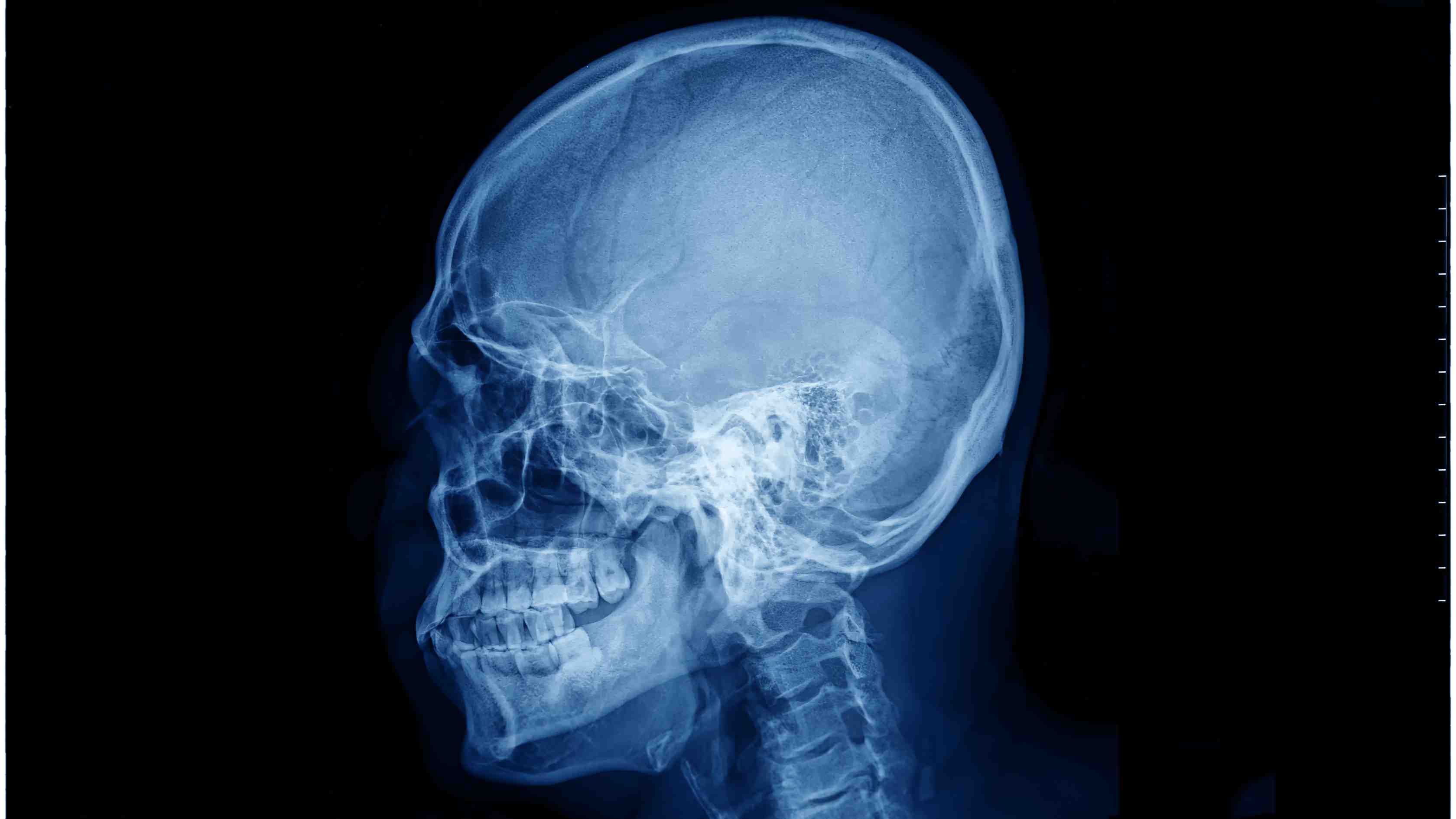
How Is Cephalometric Imaging Used in Dentistry?
Cephalometric imaging is an essential tool in modern dentistry, particularly for orthodontics and maxillofacial treatments. Its primary purpose is to evaluate the structural relationships of the jaw, teeth, and facial bones, allowing dentists to design treatments with greater precision.
Orthodontic practitioners often rely on lateral cephalometric radiographs to determine the best course of action for correcting misaligned teeth or bite problems. These images provide critical data for cephalometric analysis, such as measurements of jaw angles, tooth positioning, and growth predictions. They also help identify issues like overbites, underbites, and crossbites, making them an integral part of planning orthodontic appliances like braces or retainers.
In addition to orthodontics, cephalometric x-rays are used to evaluate airway obstructions, diagnose temporomandibular joint disorders (TMJ), and plan corrective jaw surgeries. The detailed view provided by this technology ensures that treatment plans are accurate, safe, and effective.The Role of Digital Cephalometric Analysis in Diagnosis
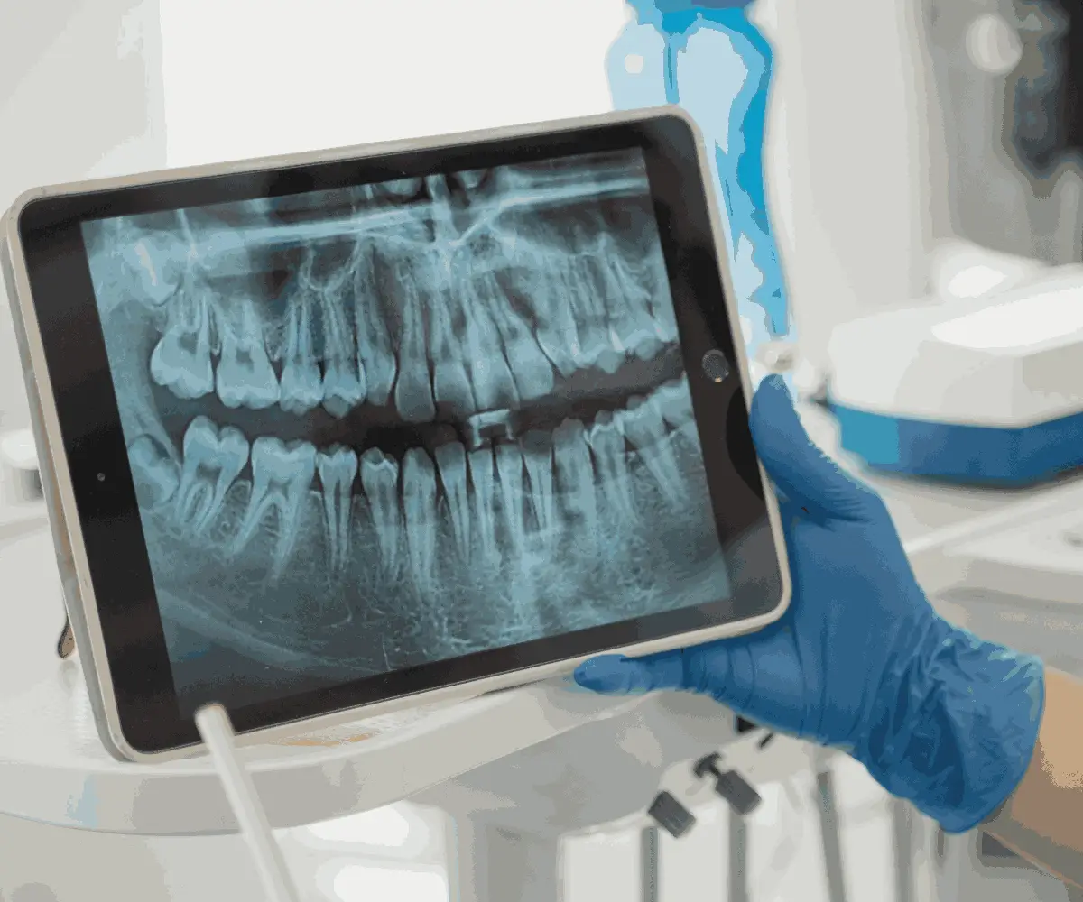
Digital cephalometric analysis revolutionizes the way orthodontists and dentists evaluate their patients. This advanced approach uses digital imaging software to enhance the accuracy and efficiency of traditional cephalometric radiography.
With digital cephalometric imaging, practitioners can quickly measure facial structures and analyze jaw-to-teeth relationships. The software allows for automated calculations of angles and proportions, eliminating human error and providing a higher level of precision. This is especially beneficial for predicting growth patterns and determining the ideal orthodontic interventions.
Another advantage of digital systems is their ability to store, retrieve, and share images effortlessly. This not only facilitates collaboration among dental professionals but also improves patient education, as they can clearly visualize the issues being addressed. Overall, digital cephalometric analysis supports better diagnostic outcomes and enhances the patient experience.
Cephalometric X-Ray and Cephalometric Radiograph Differences
The terms cephalometric x-ray and cephalometric radiograph are often used interchangeably, but they refer to the same diagnostic imaging technique. Both capture a lateral view of the craniofacial region to assess the alignment of the jaw, teeth, and skull.
However, when comparing cephalometric imaging to other radiographic methods, its unique features stand out. Unlike panoramic x-rays, which provide a wide-angle view of the entire mouth, cephalometric radiographs focus specifically on the side profile of the head. This detailed perspective is crucial for evaluating jaw alignment and planning orthodontic treatments.
Advances in digital cephalometric imaging further differentiate this technique by offering higher resolution and faster processing times. These improvements make digital cephalometric analysis a more efficient and accurate option for modern dental practices.How Does Digital Cephalometric Imaging Work?
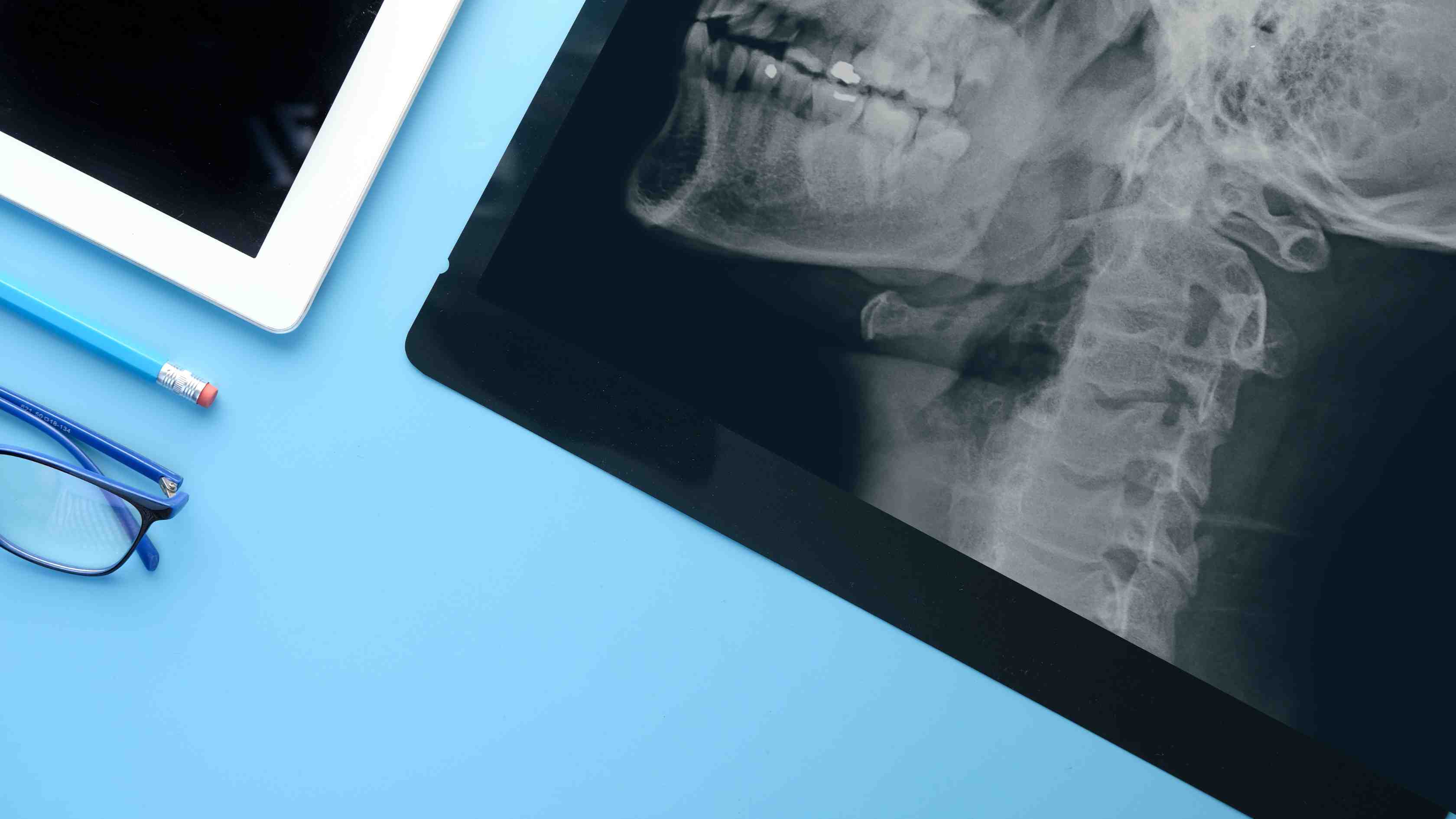
Digital cephalometric imaging works by capturing high-resolution, two-dimensional images of the head using advanced digital sensors. Unlike traditional film-based systems, digital imaging converts x-rays into digital files, enabling instant access and manipulation.
The process begins with the patient positioned in front of a cephalostat, a device that holds the head still. A lateral x-ray is taken, showing the side profile of the skull, jaw, and teeth. Once the image is captured, it is processed through specialized software for cephalometric analysis. Dentists can then measure angles, assess relationships, and identify any abnormalities.
The benefits of digital cephalometric imaging include reduced radiation exposure, faster results, and more precise measurements. These advancements significantly enhance the diagnostic process, ensuring accurate treatment planning and improved patient outcomes.
.jpg)
Common Uses of Cephalometric Imaging in Dentistry
Cephalometric imaging has numerous applications in dentistry, ranging from orthodontic treatment planning to complex surgical interventions. Here are some of its most common uses:
- Orthodontic treatment planning: Helps determine the ideal positioning of teeth and jaw alignment for braces or other corrective devices.
- Growth monitoring: Tracks jaw development in children and adolescents, ensuring timely interventions.
- Airway analysis: Assesses potential obstructions related to sleep apnea or other breathing issues.
- Surgical planning: Provides detailed measurements for jaw surgeries or facial reconstruction procedures.
- TMJ evaluation: Identifies issues with the temporomandibular joint that may cause pain or dysfunction.
With advancements in AI for dental technologies, cephalometric imaging is becoming even more precise and efficient. With advancements in AI for dental technologies, cephalometric imaging is becoming even more precise and efficient. You can visit our website and contact us to learn more about how AI in dental imaging transforms dentistry.
In summary, cephalometric radiography remains a cornerstone of orthodontic and dental diagnostics, providing invaluable data for effective and personalized treatment plans.

 Contact Us