What is Panoramic X-Ray?
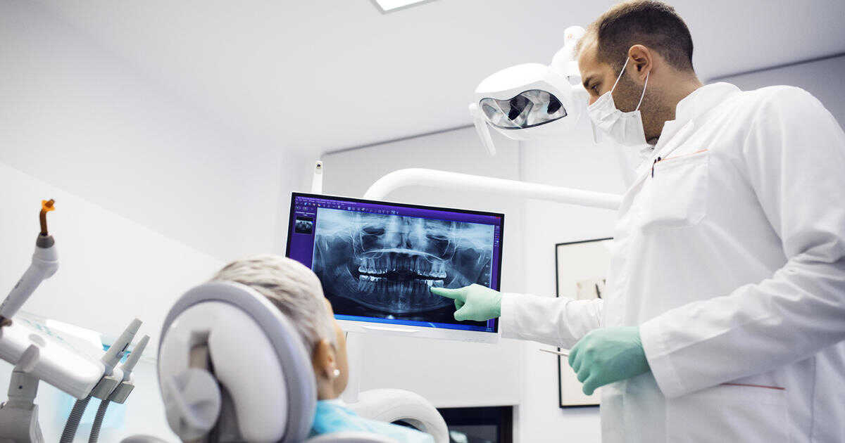
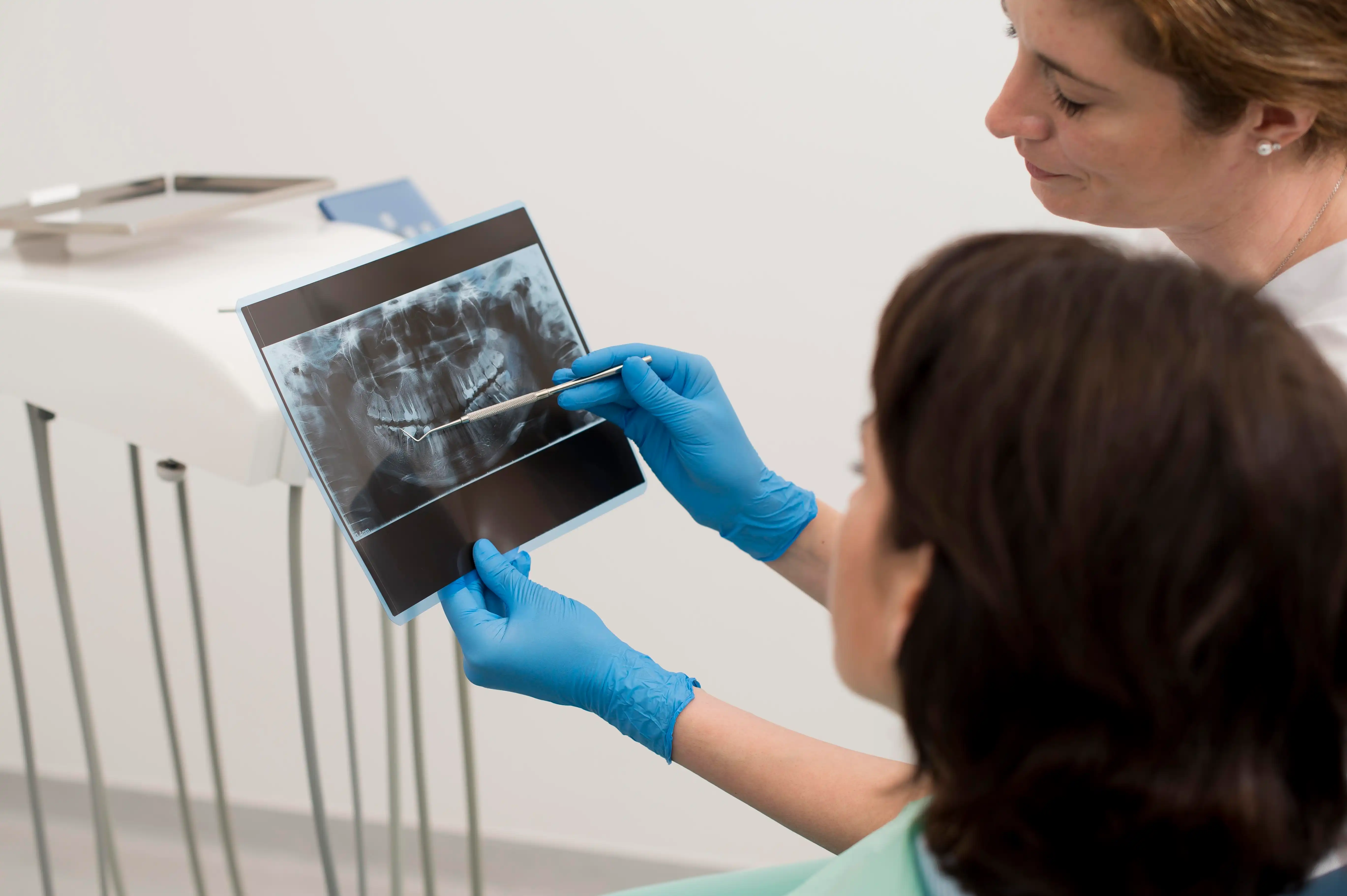
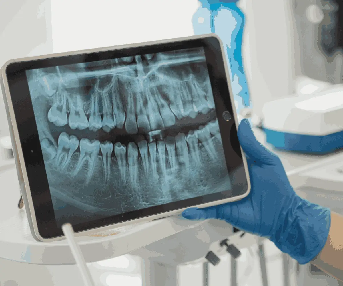
Panoramic dental x-ray is a type of dental imaging that captures the entire mouth in a single, comprehensive image. Unlike traditional intraoral x-rays that focus on individual teeth, panoramic dental x-rays offer a broader view that includes the teeth, jaws, and surrounding tissues. This advanced imaging method is essential in detecting various dental conditions that may not be visible with other types of x-rays.
Panoramic x-rays are particularly useful for diagnosing dental abnormalities, such as impacted teeth, jawbone irregularities, and infections. The panoramic teeth x-ray technology allows dentists to get a full view of the mouth without requiring multiple images. It is commonly used for treatment planning in complex dental procedures like implants, orthodontics, and extractions.
Panoramic x-ray machines are designed to rotate around the patient’s head, creating a detailed image of the upper and lower jaws. These machines use low radiation levels, which makes the procedure safer compared to other types of x-rays. The imaging is completed quickly, typically taking less than 30 seconds, providing a convenient and efficient diagnostic tool for both patients and dentists.
Understanding Panoramic Dental X-Ray Technology
The panoramic dental x-ray technology is a cutting-edge tool that provides dentists with a complete view of the mouth, including teeth, jawbones, and other vital structures. Unlike traditional x-rays, which require multiple images to cover different areas of the mouth, panoramic x-rays capture everything in one single image. This provides a more efficient way to assess a patient’s overall oral health.
During the procedure, the panoramic x-ray machine rotates around the patient’s head while they bite down on a small piece to stabilize their mouth. The x-ray machine’s tube moves in a semicircle, capturing a comprehensive image without the need for the patient to open wide or reposition several times.
One of the primary benefits of this technology is that it uses minimal radiation compared to conventional dental x-rays. This is a significant advantage for both safety and diagnostic accuracy. Panoramic dental x-rays are especially useful for detecting impacted teeth, assessing jawbone health, and planning treatments like dental implants or extractions. The clear, wide-field view allows for early detection of dental issues that might otherwise be missed, ensuring better treatment outcomes.How Do Panoramic X-Rays Work?
Panoramic x-ray technology works by rotating around the patient’s head to capture a full view of the oral structures, including teeth, jaws, and supporting bone structures. The process involves the patient standing or sitting in front of the x-ray machine while biting on a plastic piece to stabilize their mouth. The x-ray tube moves in a semicircular motion around the head, capturing images from multiple angles to provide a complete picture of the mouth.
During the scan, the machine takes a single image of the entire mouth, which helps dentists detect dental issues such as impacted wisdom teeth, jawbone abnormalities, and infections. The comprehensive view provided by a panoramic x-ray makes it easier for dental professionals to identify problems that might be missed with traditional intraoral x-rays. It is also highly beneficial for planning complex dental treatments, such as orthodontics and implants, by providing detailed information about the patient’s bone structure.
The key feature of a panoramic dental x-ray is its ability to minimize radiation exposure. Unlike conventional x-rays that require multiple images, a single panoramic scan captures everything in one go, reducing the total amount of radiation absorbed by the patient. This makes it a safer, faster, and more comfortable diagnostic tool for both patients and dentists.
How Panoramic X-Rays Differ from Regular Dental X-Rays
Panoramic dental x-rays and regular intraoral x-rays serve different purposes in dentistry. While both are important diagnostic tools, there are several key differences between them. Below are some of the main distinctions:
• Field of View: A panoramic x-ray captures the entire mouth in a single image, including both jaws, teeth, and surrounding tissues. In contrast, regular x-rays focus on specific teeth or small sections of the mouth.
• Radiation Exposure: Panoramic x-rays typically involve lower radiation exposure since they require only one image, while regular dental x-rays may need multiple images to cover the same area.
• Diagnostic Scope: Panoramic teeth x-rays are ideal for detecting broader dental issues such as jaw abnormalities, impacted teeth, and bone loss, whereas regular x-rays are more suited for detailed images of specific teeth or areas with suspected decay.
• Procedure Time: A panoramic x-ray is faster, taking about 20-30 seconds to complete, while regular x-rays may take longer, especially if multiple angles are needed.
• Comfort: Since a panoramic x-ray requires minimal patient movement and no intraoral sensors, it is often more comfortable, especially for patients with sensitive gag reflexes.
By using a panoramic dental x-ray, dentists can evaluate the entire oral cavity at once, which is especially helpful for comprehensive treatment planning.
Benefits of Using Panoramic X-Ray in Dentistry
Panoramic x-rays offer numerous advantages in the field of dentistry. These benefits enhance both the diagnostic process and the treatment planning for patients. Below are some of the key benefits of panoramic dental x-rays:
• Comprehensive View: Provides a complete image of the entire mouth, including both jaws, teeth, and surrounding tissues, which is essential for accurate diagnosis.
• Early Detection: Helps identify dental issues such as cavities, gum disease, and impacted teeth before they become more serious.
• Efficient Treatment Planning: Assists dentists in creating detailed treatment plans for procedures like dental implants, root canals, and extractions by offering a clear view of the jawbone and teeth alignment.
• Minimized Radiation Exposure: Uses lower radiation levels compared to multiple intraoral x-rays, ensuring patient safety during the imaging process.
• Quick and Painless: The procedure is non-invasive and can be completed in a matter of seconds, making it a convenient option for patients.
Panoramic x-rays are a crucial tool in modern dentistry, offering both diagnostic and procedural advantages that improve patient care and treatment outcomes.

Why Do Dentists Use Panoramic Dental X-Rays?
Dentists use panoramic dental x-rays because they offer a complete view of the patient’s oral cavity in one image. This type of imaging is particularly beneficial for detecting conditions that may not be visible during a routine dental exam. For example, panoramic x-rays can reveal issues such as impacted wisdom teeth, jawbone irregularities, tumors, and infections, which are often difficult to detect with traditional x-rays.
Another reason dentists prefer panoramic x-rays is that they provide a non-invasive and quick method to assess the overall oral health of a patient. This imaging tool is also valuable for treatment planning, especially for complex procedures like dental implants, orthodontics, or extractions. By having a complete view of the mouth, dentists can develop more accurate and personalized treatment plans.
Overall, the ability to detect a wide range of oral issues, combined with minimal discomfort for the patient, makes panoramic dental x-rays an essential tool in modern dentistry.
Diagnosing Tooth and Jaw Issues with Panoramic X-Rays
Panoramic dental x-rays are a powerful diagnostic tool that help dentists identify a variety of tooth and jaw issues. One of the main advantages of this imaging technique is that it provides a broad view of both the upper and lower jaws, enabling the dentist to detect problems that may not be visible with regular dental exams or intraoral x-rays.
With a panoramic x-ray, dentists can diagnose conditions such as:
• Jawbone abnormalities: Including fractures, cysts, or tumors that could affect a patient’s oral health.
• Impacted teeth: Particularly useful for identifying wisdom teeth that have not yet emerged or are stuck in the jawbone.
• Dental infections: These can be spotted in areas around the teeth or gums that may not show symptoms initially.
• Tooth misalignment or crowding: Helps in planning orthodontic treatments by showing the position of all teeth in relation to each other.
By using panoramic x-rays, dentists can detect and diagnose these issues early, allowing them to intervene and provide the necessary treatment before the conditions worsen.How Panoramic X-Rays Help in Planning Dental Treatments
Before performing complex dental procedures, dentists often rely on panoramic dental x-rays to assess the overall condition of the patient’s mouth. This imaging tool offers a complete picture of both the teeth and jawbones, which is crucial for planning treatments like implants, root canals, or extractions.
With a panoramic x-ray, dentists can evaluate the bone density and structure in the jaw, ensuring that there is enough support for dental implants. For root canals or extractions, it helps identify the exact position of the tooth roots and any surrounding infections or abnormalities that may complicate the procedure. Additionally, panoramic x-rays are helpful in orthodontic treatment planning, as they provide a clear view of how the teeth are aligned.
Overall, panoramic x-rays aid in creating accurate and personalized treatment plans, improving the likelihood of successful outcomes in dental procedures.
Identifying Wisdom Teeth and Impacted Teeth with Panoramic X-Rays
One of the primary uses of panoramic dental x-rays is to identify the presence and position of wisdom teeth, as well as any impacted teeth. Impacted teeth occur when a tooth does not fully emerge from the gums or gets stuck in the jawbone. This can lead to infections, misalignment, or pain if not treated.
Using a panoramic x-ray, dentists can see the exact location of wisdom teeth, even if they have not yet broken through the gums. This information is essential for planning extractions or other treatments to prevent future complications. In addition to wisdom teeth, panoramic x-rays help in identifying other impacted teeth, which may cause crowding or misalignment.
By spotting these issues early, dentists can recommend the most appropriate treatment, reducing the risk of pain, infections, and long-term dental problems.
Detecting Cavities and Dental Decay Through Panoramic X-Rays
While traditional x-rays are commonly used to detect cavities, panoramic x-rays also play a role in identifying dental decay, especially in hard-to-see areas. These x-rays provide a comprehensive view of the mouth, making it easier to spot cavities between teeth, under fillings, or in areas that might be hidden from a typical dental exam.
Panoramic x-rays are particularly useful in cases where multiple teeth are affected by decay, allowing dentists to plan comprehensive treatments to restore the patient’s oral health. Early detection of cavities through a panoramic teeth x-ray helps prevent further damage to the tooth structure and reduces the need for more invasive procedures like root canals or extractions.
By using panoramic dental x-rays, dentists can take a proactive approach to treating cavities and decay before they cause more significant oral health problems.
How Panoramic X-Rays Help in Planning Implant Placements
For patients considering dental implants, panoramic dental x-rays are an essential part of the planning process. Implants require precise placement into the jawbone, and a panoramic x-ray provides critical information about the bone structure, density, and available space in the jaw.
By analyzing the panoramic x-ray, dentists can determine whether the patient has enough healthy bone to support an implant and where the implant should be placed for optimal results. In some cases, bone grafting may be needed to strengthen the area before implant surgery can proceed. Panoramic x-rays also help identify any nearby nerves, sinuses, or other anatomical structures that need to be avoided during surgery.
With this comprehensive information, dentists can create a detailed treatment plan, ensuring the success and longevity of the dental implants.

Benefits and Risks of Panoramic Dental X-Rays
Panoramic dental x-rays are widely used in dentistry due to their numerous advantages, but they also come with certain risks. Understanding both the benefits and potential drawbacks can help patients make informed decisions about their dental care.
Benefits of Panoramic X-Rays:
• Comprehensive imaging: One of the main advantages is that panoramic x-rays capture the entire mouth in a single image, providing a broad view of the teeth, jaws, and surrounding structures. This allows for the early detection of issues like cysts, tumors, or impacted teeth.
• Reduced radiation exposure: Compared to multiple intraoral x-rays, panoramic dental x-rays involve lower radiation exposure since only one image is taken to cover the entire mouth.
• Quick and convenient: The procedure takes only about 20-30 seconds, and the patient does not need to endure multiple sessions of imaging. It is also more comfortable, especially for patients with a sensitive gag reflex.
• Effective for treatment planning: Panoramic x-rays are invaluable for planning dental treatments like implants, orthodontics, and extractions, as they provide detailed information about the patient’s bone structure and tooth positioning.
Risks of Panoramic X-Rays:
• Radiation exposure: While the radiation dose is lower than that of traditional x-rays, there is still some radiation exposure involved. However, modern machines use minimal doses to reduce this risk.
• Limited detail in certain areas: In some cases, panoramic x-rays may not provide as much detail as intraoral x-rays, especially for detecting small cavities or detailed root structures. Additional imaging may be required for specific issues.
• Positioning sensitivity: If the patient moves during the procedure or the head is not correctly positioned, the image quality can be compromised, leading to the need for retakes.
Despite these minor risks, panoramic dental x-rays remain an essential diagnostic tool in modern dentistry, offering a balance of safety, efficiency, and comprehensive imaging for patients.
What are Some Common Uses of the Procedure?
Panoramic dental x-rays have a wide range of applications in both routine dental care and more specialized treatments. Below are some of the most common uses of this imaging procedure:
• Routine dental exams: Dentists often use panoramic x-rays during regular check-ups to get a full picture of the patient’s oral health. This allows for early detection of issues that might not be visible during a physical exam.
• Orthodontic assessments: Panoramic x-rays are frequently used to evaluate the alignment of the teeth and jaws before starting orthodontic treatments like braces. This helps in identifying tooth crowding, impacted teeth, or misaligned jaws.
• Wisdom teeth extraction: One of the most common uses of panoramic x-rays is to assess the position and development of wisdom teeth. If the wisdom teeth are impacted or misaligned, panoramic imaging provides the necessary information for planning an extraction.
• Implant planning: Panoramic dental x-rays are crucial in determining whether a patient has enough bone density to support dental implants and identifying the optimal placement sites.
• Jawbone evaluation: Panoramic imaging helps detect jawbone abnormalities, fractures, or infections that could affect a patient’s overall oral health.
By using panoramic x-rays in these scenarios, dentists can ensure that they have a complete understanding of the patient’s dental health and provide more effective and tailored treatments.
What to Expect During a Panoramic X-Ray Procedure
During a panoramic dental x-ray procedure, you will be positioned in front of the panoramic x-ray machine, which will capture a single, comprehensive image of your mouth. The process is quick and painless, typically taking less than 30 seconds to complete. Here’s what to expect during the procedure:
1. Positioning: You will either stand or sit in front of the machine, depending on its design. The technician will ask you to bite down on a small plastic piece to keep your mouth steady and in the correct position.
2. Stabilization: Your head will be gently secured to prevent movement during the scan. This ensures that the resulting image is clear and accurate.
3. Rotation of the machine: Once you are in position, the panoramic x-ray machine will rotate around your head in a smooth motion, taking pictures from various angles to create a complete image of your teeth, jaws, and surrounding structures.
4. Completion: The scan is completed in about 20-30 seconds, and the resulting image is immediately available for the dentist to review.
The procedure is non-invasive, and the amount of radiation exposure is minimal. This makes panoramic dental x-rays a safe and effective option for obtaining detailed images of the mouth.
Process of Rotating Around Your Head for Panoramic X-Ray
During a panoramic x-ray scan, the machine moves in a semicircular motion around your head to capture a full image of your oral structures. This smooth rotation is key to ensuring that the entire mouth, including the teeth, jawbones, and surrounding tissues, is included in the image.
The process begins by positioning the patient securely in front of the machine. The patient bites down on a small piece to stabilize their mouth, while their head is held in place to prevent movement. As the panoramic x-ray machine rotates, it captures images from multiple angles, allowing for a comprehensive view of both the upper and lower jaws.
One of the benefits of this rotating technique is that it reduces the need for multiple images, as everything is captured in one pass. This not only speeds up the process but also minimizes radiation exposure. The rotation also ensures that the x-ray includes all necessar
FAQ About Panoramic Dental X-Ray
Here are some frequently asked questions about panoramic dental x-rays:
Q: Why are panoramic x-rays used in dentistry?
A: Panoramic x-rays are valuable diagnostic tools that provide a comprehensive view of the oral and maxillofacial area, helping dentists detect issues such as impacted teeth, jaw disorders, and bone abnormalities.
Q: Are panoramic x-rays safe?
A: Yes, panoramic x-rays are considered safe and emit very low levels of radiation. Dentists take necessary precautions to minimize radiation exposure during the imaging process.
Q: How often should someone have a panoramic x-ray taken?
A: The frequency of panoramic x-rays varies depending on an individual's oral health needs and risk factors. Dentists typically recommend them every 3-5 years for most patients.
Q: What information can dentists gather from a panoramic x-ray?
A: Dentists can assess the positions of teeth, look for signs of decay, evaluate bone health, monitor growth and development, and detect any abnormalities in the oral and maxillofacial region using a panoramic x-ray.
If you found the topic Dental Imaging Insights: Panoramic X-Ray in Dentistry interesting, be sure to check out our article on Dental Imaging Essentials: A Comprehensive Guide for Radiologists.


 Contact Us

